It starts with a kiss: the fluid mechanics of sample preparation in Cryo-Electron Microscopy
- Academic lead
- Nikil Kapur, School of Mechanical Engineering, n.kapur@leeds.ac.uk
- Co-supervisor(s)
- Rob Kay, School of Mechanical Engineering, r.w.kay@leeds.ac.uk, Stephen Muench, Faculty of Biological Sciences, s.p.muench@leeds.ac.uk, Louie Aspinall, Faculty of Biological Sciences, l.p.aspinall@leeds.ac.uk
- Project themes
- Biomedical Flows
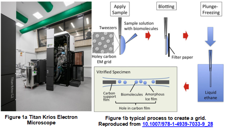
Cryo-electron microscopes (cryo-EM) have proved vital in developing understanding of biological molecules such as proteins and viruses, and then going on to support the development of modern medicines to treat (for example) cancer and viral infections [1]. There has been a huge investment in technology and modern microscopes (such as the one in Astbury that is part of this project (fig 1a)) which cost in the region of £20M and offer an unprecedented level of performance. However, the preparation of the sample which is imaged in the microscope is now a recognised bottleneck [2] and is limiting the use of cryo-EM more broadly.
The process to create the ‘sample grid’ (essentially the structure onto which the sample is placed in a way to allow imaging) is crude and does indeed start with a kiss! A drop of sample is touched onto the grid, then it is blotted to remove most of it, then it is plunged into liquid ethane to form vitreous ice which is transparent to electrons (fig 1b).
The whole process has largely been artisan developed and remains unchanged since the advent of cryo-EM. But there is so much fluid mechanics that could be used to understand the science of this process and drive innovative thinking. The work will draw on science around droplets, transport of particles, porous media and drying and evaporation and will see you develop your skills across experiments, analysis and modelling.