Multi‐phase CFD modelling of biological tissue flow with applications in liver surgery
- Academic lead
- Sven Van Loo (Physics and Astronomy)
- Industrial lead
- Gunnar Schuetz, Bayer AG
- Co-supervisor(s)
- Steven Sourbron (Medicine and Health), Amirul Khan (Civil Engineering)
- Project themes
- Microflows & heat transfer
Computational Fluid Dynamics has been used extensively in medicine to model the flow of blood in large arteries and veins and in the heart, but our understanding of the spatial propagation of body fluids on the microscopic level is more limited. The purpose of this PhD project is to enable the study of these processes by developing multi‐phase CFD models for flow, diffusion and permeability in healthy and diseased biological tissues. We will apply methods and concepts from porous media studies and use those to develop models for body tissues that are able to describe 4D dynamic data obtained by Magnetic Resonance Imaging (MRI) and indicator‐dilution experiments. These methods will then be deployed in clinical studies to assess how they can improve survival of patients with liver cancer through better predictions of surgical risk. The project involves a collaboration between academic supervisors from various backgrounds (Medical Imaging, Physics and Civil Engineering), and an industrial sponsor in a pharmaceutical company (Bayer). The student will have a unique opportunity to gain in‐depth knowledge of the theoretical and numerical basis of multi‐phase CFD modelling and data assimilation techniques, but also about their applications in medicine ‐ a novel application area with major growth potential for CFD. The industrial sponsor will provide an opportunity for a temporary placement in Berlin where the student will learn how to translate research ideas into patient benefit.
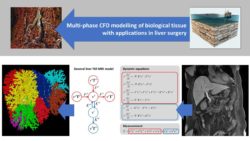
Figure 1. This project involves the application of porous media theory to body tissue (top row) and will aim to develop spatiotemporal models for complex body tissues (bottom row, middle) that can accurate describe 4D dynamic MRI data (bottom row, right), and use these to extract currently invisible information on tissue microflow (bottom row, left). We expect the new information to be useful in improving the planning of liver resections for treatment of cancer, as well as many other application areas.